Globally, there are over 50 species of deer, most of which are found in Asia, although many species have been introduced outside of their original habitats as game animals.1 Deer are cervids (family Cervidae), a group of animals belonging to the order Artiodactyla. They have a four-chambered stomach, and are cud-chewing herbivores (ruminants).
The Eld's deer (Rucervus eldii) is native to areas of Southeast Asia. So named because of their discovery by Lt. Percy Eld in the Manipur Valley of India in 1838, there are three recognized subspecies:
- Rucervus eldii eldii (native to Manipur),
- Rucervus eldii thamin (native to Burma/Myanmar), and
- Rucervus eldii siamensis, (native to Thailand, Annam, and Hainan island).
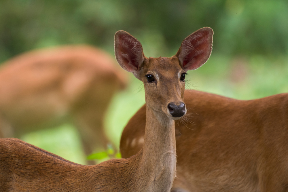
The Eld’s deer is a large deer that is considered striking and elegant in appearance. They are similar in size to white-tailed deer, but differ somewhat in appearance. Their rough coats change color with the season. In summer, they are reddish-brown, and dark brown in winter. Stags often have darker coloring than hinds (females) and have a thick mane of long hair around the neck. Their legs are long and thin, and they have slender bodies with a large head and ears.1
Eld’s deer stags have large lyre-shaped antlers; these sweep back in a curve of about 40 inches in length, with one smaller tine growing toward the front of the head. The antlers are shed every year and reach their largest size during the breeding season.2 Male Eld’s deer grow to about 71 inches in length and weigh from 276 to 386 pounds. They are taller and larger than the hinds, which stand about 60 inches tall.
Eld’s deer inhabit suitable forest habitats, lowland valleys and plains in their native ranges. Today, they occur in a number of protected areas throughout these areas and have been introduced to numerous countries as game animals, including the United States.2
Eld’s deer are associated with areas that are seasonally burned, and are fond of eating new grasses as they emerge after fires. Their diet consists largely of grasses, fruits, herbaceous and wetland plants. They are also known to graze and to browse on cultivated crops such as rice, lentils, corn and peas. On ranches and reserves, Eld’s deer are typically fed a low-protein herbivore diet and alfalfa hay.1,3
Eld’s deer hinds can begin reproducing at two years of age and typically continue to do so until they are 10 years of age. They begin estrus in the late winter or early spring and have a long period of ovarian activity (225 to 342 days), during which they average 10 to 17 estrous cycles. After they have mated, the females enter anestrus, which usually occurs in the autumn months.2
The Eld’s deer is primarily nocturnal. Throughout most of the year, stags tend to be loners, except in the spring when mating commences. Females are generally found alone or in pairs with their young. They remain in close association with their fawns and other female-fawn pairs. Larger groups are often formed when males join groups of females prior to the breeding season, and groups of up to 20 animals are common.2,3
How Shock Arises in Eld’s Deer
Shock is a critical condition that comes about due to a sudden drop in blood flow throughout an animal’s body. It can be the result of a wide variety of conditions or circumstances, including extreme physical stress, trauma, disease, heatstroke, blood loss, allergic reactions or severe infection. When a deer is suffering from shock, its organs are not receiving an adequate amount of blood or oxygen. If untreated, this condition can lead to permanent organ damage or death.
Unfortunately, the processes surrounding capture and/or chemical immobilization can include extreme physical stress and/or trauma sufficient to induce shock in Eld’s deer and other animals. The degree of risk is dependent upon factors such as species, sex, age, overall health, environmental factors, length of immobilization, the degree of stress involved in the capture/immobilization event itself, the specific chemical agents involved in immobilizing the animal and others. Cervids are known to be susceptible to a variety of complication during chemical immobilization, including shock.
There are three categories of shock, which include:
Circulatory Shock. This occurs when there is a significant decrease in effective circulating blood volume. Circulatory shock is further divided into the three subcategories of cardiogenic, hypovolemic and distributive shock. Cardiogenic shock occurs when the circulating volume of blood decreases despite normal or increased blood volume. Hypovolemic shock occurs when blood volume is decreased through hemorrhage, third space fluid distribution, or dehydration. Distributive shock occurs when an animal’s body is unable to maintain the vasoconstriction of blood vessels.5
The last two categories of shock are hypoxic shock and metabolic shock. Hypoxic shock results from impaired oxygen delivery to cells, while metabolic shock involves cells that have become unable to utilize oxygen for energy production.5,6The types of shock being discussed here are the subcategories of circulatory shock and hypoxic shock, which are the most likely to be brought on due to capture and/or chemical immobilization events.
The Mechanics of Shock in Eld’s Deer
Chemical immobilization is a risky proposition in Eld’s deer and other cervids even under the best of circumstances. Almost all of the drugs that produce anesthesia compromise cardiovascular stability by producing dose-dependent impairment of cardiac function, vascular reactivity and autoregulatory responses.6 Hemoglobin is found within red blood cells and carries oxygen to tissues. Normally, the amount of oxygen delivered to cells is 2 to 4 times the amount required, which ensures an adequate supply.2 However, if tissues are not adequately perfused with blood, oxygen fails to get to the cells, regardless of the oxygen content in the blood.5
Any significant change in the mean arterial pressure (MAP) can trigger changes in heart rate.6,7 An increase in MAP also causes bradycardia and vasodilation, while a decrease produces tachycardia and vasoconstriction.5,6 While anesthesia-related depression of cardiac function and arterial vasodilation are adverse effects that are well-recognized as contributing to anesthetic risk, far less emphasis is usually placed on effects impacting venous physiology and venous return.4
Approximately 70% of an animal’s total blood volume is represented by venous circulation, and this is a chief contributor to stroke volume and cardiac output.5 Vasodilation is the primary cause of hypovolemia produced by anesthetic drugs. It is often associated with increased venous compliance, decreased venous return, and reduced response to vasoactive substances.6 Depending on things like patient status and monitoring, a state of relative hypovolemia can remain clinically undetected for long periods of time.5-7
Diagnosis and Treatment of Shock in Eld’s Deer
The clinical signs of shock in deer can include any combination of the following symptoms:
- Unresponsiveness
- Hypothermia
- Tachycardia
- Bradycardia
- Tachypnea
- Bradypnea
- Marked hypotension
- Cyanosis
- Orthopnea
The treatment of shock in Eld’s deer must primarily focus on increasing oxygen delivery to the tissues. This can be accomplished by providing supplemental oxygen, increasing effective circulating volume, increasing hemoglobin concentration and increasing cardiac output with stimulants.5,7 An intravenous catheter should be placed for vascular access if possible. If venous access cannot be established, an intraosseous catheter can be placed. Oxygen supplementation, when available, will also provide benefits to the deer suffering from shock. This can be accomplished via flow-by oxygen, mask, nasal cannulas or an oxygen cage.6
Fluid Therapy for Shock in Eld’s Deer
Lactated Ringer’s solution, Normosol-R, and Plasma-Lyte are the preferred choices for fluid therapy in treating shock in Eld’s deer, as these have been shown to cause fewer complications as well as decrease the risk of mortality5 as compared to other options. Hypertonic saline is also a popular option for fluid therapy (as this increases vascular volume). Hypertonic saline increases plasma osmolarity, pulling water into the vascular space from the interstitial space, thereby expanding plasma volume. It should be noted that hypertonic saline does have unwanted side effects, such as a transient, dose-dependent increase in sodium and chloride.6,7
Blood products are an important adjunct for the treatment of shock in Eld’s deer. In healthy animals, anemia can be well-tolerated with oxygen delivery being maintained. In patients with trauma and acute loss of blood volume however, the associated stressors can contribute to decreased oxygen delivery.7
1animaldiversity.org.
2nationalzoo.si.edu.
3animalia.bio.
4todaysveterinarynurse.com.
5Noel-Morgan, J., Muir, W. (2018) Anesthesia-Associated Relative Hypovolemia: Mechanisms, Monitoring, and Treatment Considerations. Frontiers in Veterinary Science, Vol. 5 (53).
6Haller G, Laroche T, Clergue F. Morbidity in anaesthesia: today and tomorrow. Best Pract Res Clin Anaesthesiology (2011) 25(2):123–32.
7Steadman J, Catalani B, Sharp CR, Cooper L. Life-threatening perioperative anesthetic complications: major issues surrounding perioperative morbidity and mortality. Trauma Surg Acute Care Open (2017).


