The Eld’s deer (Rucervus eldii) is a tropical deer that is native to the lowland forests of a region known as the Indo-Burma Biodiversity Hotspot, which includes forested areas, lowland valleys and plains of southeast Asia.1 Today, Eld’s deer occur in several protected areas throughout their native range and have been introduced into numerous other countries as game animals.
Three subspecies of Eld’s deer are currently recognized:
- The Sangai (Rucervus eldii eldii) of northeast India.
- The Thamin (Rucervus eldii thamin) of Myanmar and western Thailand.
- The Siamese Eld’s Deer (Rucervus eldii siamensis) of Indochina and China’s Hainan Island.2
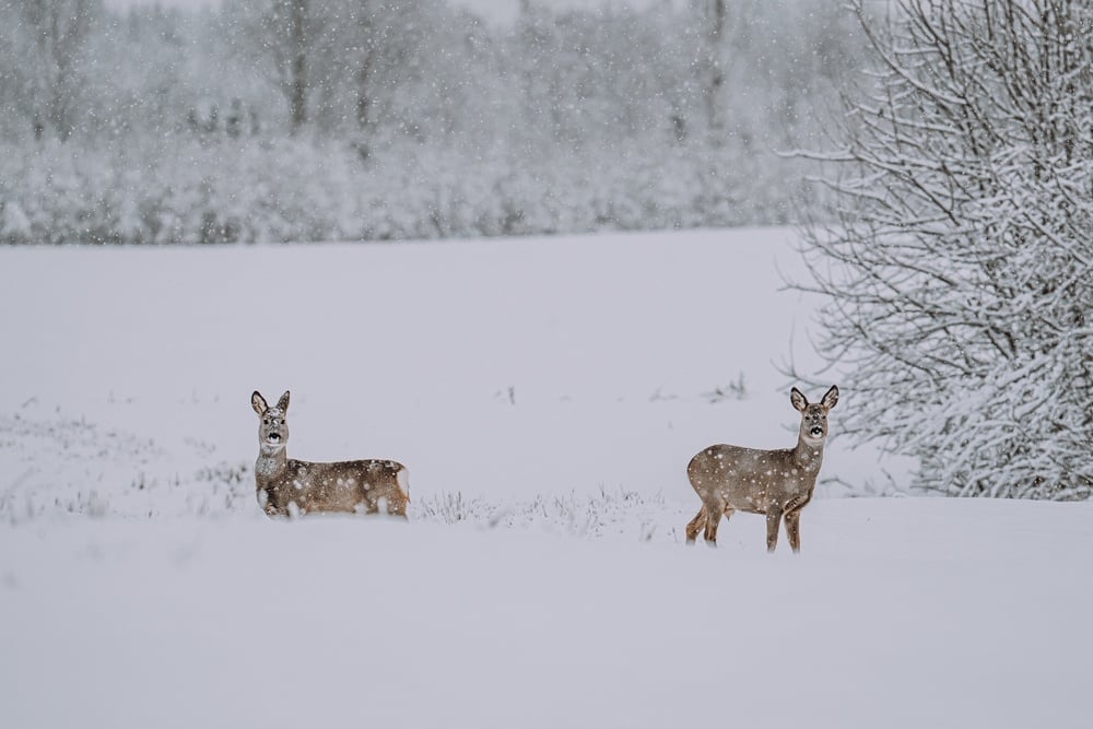
The Eld’s deer is a large deer that is similar in size to white-tailed deer, but differs in body type. Their legs are long and thin, and they have slender bodies with a large head and ears. Their rough coats change color with the season. In summer, they are reddish-brown, and dark brown in winter. Stags often have darker coloring than hinds (females) and have a thick mane of long hair around the neck.1
Eld’s deer are known to graze and browse on wild fruits and cultivated crops from nearby fields. Rucervus eldii thaminhas been reported to eat the fruits of various woody plant species. They also eat forbs and grasses in these areas.3 Their feeding may vary seasonally with food availability and with reproductive considerations; during rut, males often experience a decline in body weight, probably due to a decrease in their food intake and an increase in activity.
While Eld’s deer are usually solitary, they sometimes form large groups or herds as an anti-predator adaptation. These larger groups decrease the risk of predation, both by increasing the chance that a predator will target other animals rather than a lone individual, and by the increased vigilance for predators which can be provided by all members of the group.2The most common predators of Eld’s deer are tigers and leopards. Jackals and feral dogs occasionally hunt these deer, and poaching by humans is a serious problem to Eld's deer populations.1
Frostbite Risks and Phases of Frostbite
The chemical immobilization of Eld’s deer can require extended periods of immobility in captured animals. While hypothermia is an inherent risk to any animal undergoing chemical immobilization regardless of ambient temperature, frostbite is an even greater risk during the winter months. Frostbite is a cold-related injury in which the tissues of the body begin to freeze. It can affect any part of the body that is exposed to extreme cold for an extended period of time. This period of time is reduced as the relative temperature drops. When subjected to cold, blood vessels throughout the body constrict to preserve heat; this means that the body biochemically prioritizes keeping its core warm over the extremities. This is why they are more at risk for frostbite.
With frostnip (early-stage frostbite or the near-freezing of tissues), the skin becomes red, cold to the touch and may begin to go numb. In these cases, if the skin is warmed soon enough after exposure, there is usually no permanent damage. Continued exposure to cold can result in superficial frostbite.
In cases of superficial frostbite, ice crystals begin to form within the skin as it freezes. This can cause permanent damage to the tissue affected. At this stage, the skin may appear white and fluid-filled blisters can appear. Naturally, this may be difficult to detect in wild animals, as their bodies are covered with a fur coat. In cases of deep frostbite, large blisters form, and the tissue will often turn black and hard as it necrotizes.
Frostbite is divided into four overlapping phases:
- Prefreeze
- Freeze–thaw
- Vascular stasis
- Late ischemic4
Prefreeze consists of tissue cooling with accompanying vasoconstriction and ischemia and without ice crystal formation. The freeze–thaw phase is represented by the intracellular or extracellular formation of ice crystals. In the vascular stasis phase, vessels fluctuate between constriction and dilation, and blood may leak from vessels or coagulate within them. The late ischemic phase results from progressive tissue ischemia and infarction from a cascade of events, including inflammation, vasoconstriction and emboli.4
Classifications of Frostbite in Eld’s Deer
Frostbite is classified into four degrees of injury that follow the classification schemes for thermal burn injury which are based on acute physical findings and advanced imaging after rewarming. Early stages of frostbite are to be differentiated from frostnip, which is a superficial nonfreezing cold injury associated with intense vasoconstriction on exposed skin. As indicated earlier, frostnip may precede frostbite. In these cases, ice crystals do not form within the tissue and tissue loss does not occur.5,6
The classification for the degrees of frostbite injury include:
- First-degree frostbite causes numbness and erythema. A white or yellow, firm, and slightly raised plaque develops in the area of injury.
- Second-degree frostbite injury causes superficial skin vesiculation. A clear or milky fluid will be present in superficial blisters.
- Third-degree frostbite causes deeper hemorrhagic blisters, indicating that the injury has extended into the reticular dermis and beneath the dermal vascular plexus.
- Fourth-degree frostbite extends completely through the dermis and involves the comparatively avascular subcutaneous tissues, with necrosis extending into muscle and bone.4
A variation favored by McIntosh, et. al., involves a 2-tier classification scheme:
- Superficial—Minimal anticipated tissue loss, corresponding to first- and second-degree injury.
- Deep—Anticipated tissue loss corresponding to third- and fourth-degree injury.5
It should be noted that the severity of frostbite may vary within a single extremity.
Prevention of Frostbite in Eld’s Deer
Frostbite is not generally improved by treatment, so prevention is a far better methodology than attempting to treat affected animals. Underlying medical problems and the chemical immobilization event itself can increase risk of frostbite, so prevention must address both health-related and environmental aspects. The team in the field must ensure adequate tissue perfusion and minimize heat loss to prevent frostbite.6
Preventive measures to ensure local tissue perfusion include:
- Maintaining adequate core temperature
- Maintaining adequate body hydration
- Minimizing the effects of any known diseases that might decrease perfusion
- Covering the body and head to insulate from the cold
- Minimizing any blood flow restriction
- Using supplemental oxygen in severely hypoxic conditions4,7
Measures should also be taken to minimize exposure of the animal’s tissues to cold, such as:
- Avoid environmental conditions that predispose to frostbite if possible (e.g., below -15°C, even with low wind speeds)
- Protecting exposed skin from moisture, wind, and cold
- Avoiding perspiration or wet extremities
- Increasing insulation and skin protection
- Using chemical and/or electric warmers to maintain peripheral warmth (These should be close to body temperature before being activated and must not be placed directly against skin or constrict flow)
- Regularly checking the animal’s temperature
- Recognizing frostnip or superficial frostbite before it becomes more serious
- Minimizing duration of cold exposure4,7
Treatment of Frostbite in Eld’s Deer
Since the time that an animal’s extremities can remain numb before developing frostbite cannot be determined in a chemically-immobilized animal, any extremity at risk for frostbite (typically indicated by pale color) should be warmed.4
If a deer’s body part is frozen in the field, the frozen tissue should be protected from further damage.4,5 Then, a decision must be made whether or not to thaw the tissue. If environmental conditions are such that thawed tissue could refreeze, it is safer to keep the affected part frozen until a thawed state can be maintained. Frostbite thaws spontaneously and should be allowed to do so if rapid rewarming cannot be easily achieved.
Hypothermia frequently accompanies frostbite and causes peripheral vasoconstriction that impairs blood flow to the extremities. Mild hypothermia may be treated concurrently with frostbite injury. Moderate and severe hypothermia should be treated effectively before treating frostbite injury.7
Treatment Modalities for Frostbite in Eld’s Deer
1. Hydration
Frostbite may result in vascular stasis; thus appropriate hydration and avoidance of hypovolemia are important for frostbite recovery. Intravenous normal saline should be given to maintain normal urine output. IV fluids should optimally be warmed before infusion and infused in small, rapid boluses, as slow infusion can result in fluid cooling and even freezing as it passes through tubing. Fluid administration should be optimized to prevent clinical dehydration.6,7
2. Low Molecular Weight Dextran Treatment
Intravenous low molecular weight dextran (LMWD) decreases blood viscosity by preventing red blood cell aggregation and formation of microthrombi and can be given in the field once it has been warmed. In some animal studies, the extent of tissue necrosis was found to be significantly less than in control subjects when LMWD was used, and was more beneficial if given early.4,7
The use of LMWD has not been evaluated in combination with other treatments such as thrombolytics. LMWD should be given if the animal is not being considered for other systemic treatments, such as thrombolytic therapy.5
3. Treatment With NSAIDS
Nonsteroidal anti-inflammatory drugs (NSAIDs) block the arachidonic acid pathway and decrease production of prostaglandins and thromboxanes. These can lead to vasoconstriction, dermal ischemia, and further tissue damage.4 No studies have demonstrated that any particular anti-inflammatory agent or dosing is clearly related to outcome, however. One rabbit ear model study showed 23% tissue survival with aspirin vs 0% in the control group.6 However, aspirin theoretically blocks production of certain prostaglandins that are beneficial to wound healing.8 The authors of the rabbit ear model study recommended the use of ibuprofen instead of aspirin.
1animaldiversity.org.
2nationalzoo.si.edu.
3animalia.bio.
4McIntosh, S., et. al. Clinical Practice Guidelines for the Prevention and Treatment of Frostbite: 2019 Update. Wilderness Medical Society Clinical Practice Guidelines, Volume 30, Issue 4, Supplement S19-S32, December 01, 2019.
5McIntosh, S.E., et. al. Wilderness Medical Society practice guidelines for the prevention and treatment of frostbite: 2014 update.Wilderness Environ Med. 2014; 25: S43-S54
6Mazur P. Causes of injury in frozen and thawed cells. Fed Proc. 1965; 24: S175-S182
7Lange K., et. al. The functional pathology of frostbite and the prevention of gangrene in experimental animals and humans.Science. 1945; 102: 151-152.
8Cauchy E., et. al. Retrospective study of 70 cases of severe frostbite lesions: a proposed new classification scheme. Wilderness Environ Med. 2001; 12: 248-255.


