There are dozens of deer species that are recognized worldwide, and deer occur on all continents except Antarctica. Deer are hoofed mammals which belong to the order Artiodactyla and the family Cervidae; thus, these animals are also known as cervids. The Eld’s deer (Rucervus eldii) is a deer that is native to the lowland forests of an area known as the Indo-Burma Biodiversity Hotspot.1 This tropical species was discovered by Lt. Percy Eld in the Manipur Valley of India in 1838, from which its name is derived.
The Eld’s deer inhabits the forested areas, lowland valleys and plains of southeast Asia, and is said to avoid dense forests and coastal areas. Due to rampant hunting and habitat conversion, populations of this deer have significantly declined in their native range and are now severely fragmented. Today, Eld’s deer occur in several protected areas throughout their former range and have been introduced into numerous other countries as game animals.
There are three subspecies of Eld’s deer that are currently recognized:
- The Sangai (Rucervus eldii eldii) of northeast India. Once widespread in the Manipur Valley, this subspecies was declared extinct until it was rediscovered in Keibul Lamjao National Park.
- The Thamin (Rucervus eldii thamin) of Myanmar and western Thailand. This subspecies was once distributed widely in the central plains of Myanmar, but is now reduced to two major populations.
- The Siamese Eld’s Deer (Rucervus eldii siamensis) of Indochina and China’s Hainan Island. This subspecies once roamed Thailand, Laos, Cambodia, Vietnam and Hainan. It was thought to be extinct in the wild across Indochina, but scattered populations were rediscovered in Laos and Cambodia in 1998.2
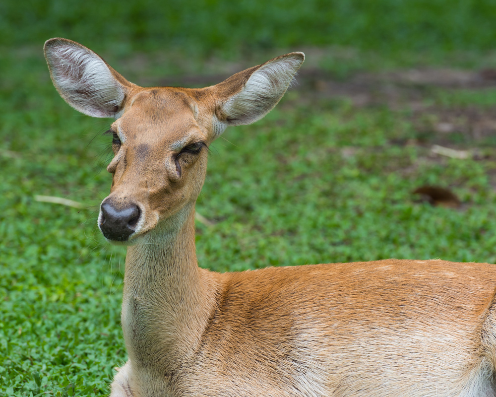
The Eld’s deer is a large deer that is considered graceful in appearance. Similar in size to white-tailed deer, Eld’s deer differ somewhat in body type. Their legs are long and thin, and they have slender bodies with a large head and ears. Their rough coats change color with the season. In summer, they are reddish-brown, and dark brown in winter. Stags often have darker coloring than hinds (females) and have a thick mane of long hair around the neck.1
Eld’s deer graze and browse on wild fruits and cultivated crops such as rice, lentils, corn, peas and rape from nearby fields. Rucervus eldii thamin has been reported to eat the fruits of various woody plant species, and also eat forbs and grasses in these areas.3 The Eld’s deer is often associated with areas that are seasonally burned, since they are often found eating new grasses as they emerge after a burn.1 Their feeding may vary seasonally with food availability and with reproductive considerations; during rut, males often experience a decline in body weight, probably due to a decrease in their food intake.
While Eld’s deer are usually solitary, they will form large groups or herds on occasion as an adaptation to avoid predators. These larger groups decrease the risk of predation, both by increasing the chance that a predator will target other animals rather than a lone individual, and by the increased vigilance for predators which can be provided by all members of the group.2 The most common predators of Eld’s deer are tigers and leopards.1
As is the case with most cervids, Eld’s deer mothers hide their fawns in underbrush immediately after birth. Females give birth during the cool-dry season when the flood waters have receded and vegetation has begun to grow, which provides the young with shelter and helps to conceal them.3 Fawns are weaned after 4 to 5 months, which allows them to have sufficient time to increase their mobility and ability to travel with the herd.1,2
Eld’s Deer and Chemical Immobilization
The field immobilization of Eld’s deer is sometimes required for medical examination, blood sample collection and animal identification, and the importance of performing such procedures for research and conservation projects is widely acknowledged.4 The need to assess the negative effects of capture is also important in order to ensure ethical standards and the validity of research results. Research in this area has revealed that the physiological and behavioral effects of capture are as important as the direct risks of injury or death of an animal.5
Several common stressors attendant to the chemical immobilization of Eld’s deer can lead to complications during or after an immobilization (sedation or anesthetic) event. The overall health of an individual animal is also a factor affecting the potential for complications during and after sedation or anesthesia.
These stressors fall into four categories:
- Physiological: Heavy exercise, hemorrhage, hyperthermia, shock, pain, infection
- Physical: Trauma/surgery, intense heat/cold
- Chemical: Hypoxemia, acid-base imbalance, anesthetic drugs
- Emotional: Anxiety, fear6
Agents involved in chemical immobilization agents are represented by the third category, although elements of the other three are often included in immobilization events. The physical stress of capture and/or attempts to escape during capture on the part of an animal certainly constitute physiological stress; surgical and even environmental conditions can bring about physical stress, and anxiety and fear are nearly always a component to some degree in a capture scenario.
Acute stress during capture events can give rise to spikes in adrenaline, cortisol levels, heart rate, blood pressure, respiration, metabolic rate, blood glucose, lactic acid and body temperature, while bringing about a decrease in pH and a redistribution of blood within the organs. The effects of capture and anesthesia can activate the fight-or-flight response, HPA-axis activation, hyperthermia, respiratory depression (hypoxemia), lactid acid build-up, acidosis; in severe cases, this can lead to neurological/myocardial dysfunction, multi-organ failure, capture myopathy and death.6
Dehydration Risks in Eld’s Deer
Dehydration may initially seem like a minor concern compared to some surgical complications, but inasmuch as it can lead directly to cardiac arrest, dehydration (a reduction of the body’s water content) is potentially very dangerous. All animals require water to ensure their bodies are working properly. It is so important that essentially all bodily functions require it to remain operative. If an animal loses more water and electrolytes (see below) than it is taking in, it will begin to dehydrate and its health will deteriorate quickly.
Electrolytes are minerals that naturally occur in all animals, and they are essential for proper health. Electrolytes are comprised of sodium, chloride, and potassium, and facilitate the movement of nutrients into cells, aid in muscle function, and help regulate nerve activities.5,6 An animal’s natural activities—breathing, urinating, and defecating, as well as simple evaporation—all cause it to lose fluids. When an animal eats and drinks, the lost water and electrolytes are replaced. If the animal’s fluid intake becomes less than what they are losing, dehydration will occur. This causes a reduction in bodily fluids that reduces blood flow and the delivery of oxygen to organs and tissues.
Understanding Dehydration in Eld’s Deer
An understanding of the distribution of fluid and water in the body is necessary in order to understand dehydration. Total body water (TBW) comprises approximately 60% of an animal’s body weight. Approximately 67% of TBW is found inside the body’s cells; this is referred to as intracellular fluid (ICF). The remaining 33% of TBW is the extracellular fluid (ECF), which comprises:
- Interstitial fluid, which bathes cells and tissues (~24% of TBW)
- Plasma, the liquid portion of blood, which constitutes most of intravascular volume (~8%–10% of TBW)
- Transcellular fluid, which comprises synovial joint fluid, cerebrospinal fluid, bile, and the fluid in the linings of the peritoneal cavity, pericardium, and pleural space (~2% of TBW)5
An approximate formula that is for the distribution of fluids in the body is the 60:40:20 rule:
- 60% of an animal’s body weight is water,
- 40% of body weight is ICF,
- and 20% of body weight is ECF.5,6
Dehydration can be brought on by hyperthermia, chronic vomiting or diarrhea, excessive urination or wound drainage. Due to the stressful nature of capture and chemical immobilization events, these have been known to bring about dehydration. In both human and veterinary practices, IV fluids are usually administered prophylactically, depending on the nature of the procedure. Veterinarians often provide fluid therapy to patients for many reasons, including correction of dehydration, expansion and support of intravascular volume, correction of electrolyte disturbances, and encouragement of appropriate redistribution of fluids that may be in the wrong compartment (e.g., peritoneal effusion).5
Each species of deer has its own anesthesia recommendation with intra-species variations of dosages because of diverse individual responses to anesthetic agents.5,6 These variations are factors for the risk of dehydration in these species, and related factors (e.g., stress, venue, individual animal and field conditions) must also be taken into account. Prior to the development of some of the newer drug formulations, some deer species were known to be very difficult to immobilize successfully.
Treating Dehydration in Eld’s Deer
In deer anesthesia, monitoring core body temperature is essential.6 During immobilization events, hydration status can be assessed using various tests. One of the easiest to perform is a skin tent test to check the turgor (moisture level) of the skin. To do this, the skin over the thorax or lumbar region is pulled away from the back. In a well-hydrated animal, the skin immediately returns to its normal resting position. If the tent formed remains standing, it is a likely indication of dehydration. If there is evidence of dehydration in a deer during a procedure, all administration of immobilizing drugs must be immediately suspended. Fluid therapy should begin in the form of lactated Ringer’s solution or 0.9% saline, IV, SQ or IP.5
Perioperative IV fluid therapy is very common in veterinary medicine and allows practitioners to restore intravascular volume, correct dehydration, and administer IV medications quickly.6 While perioperative fluid therapy under many field conditions may be impractical, fluids should always be available in the case of dehydration when chemically immobilizing Eld’s deer.
1animaldiversity.org.
2nationalzoo.si.edu.
3animalia.bio.
4Laricchiuta P, De Monte V, Campolo M, Grano F, Iarussi F, Crovace A, Staffieri F. Evaluation of a butorphanol, detomidine, and midazolam combination for immobilization of captive Nile lechwe antelopes (Kobus magaceros). J Wildl Dis. 2012 Jul;48(3):739-46.
54rivio F, Grignolio S, Sica N, Cerise S, Bassano B (2015) Assessing the Impact of Capture on Wild Animals: The Case Study of Chemical Immobilisation on Alpine Ibex. PLoS ONE 10(6): e0130957.
6Kreeger T., Arnemo, J., Raath, J. Handbook of Wildlife Chemical Immobilization, International Edition, Wildlife Pharmaceuticals, Inc., Fort Collins, CO. (2002).


