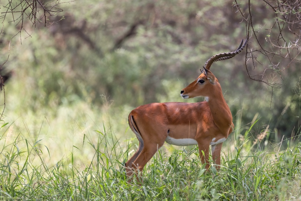
The impala, (Aepyceros melampus) is a swift, agile antelope, and the most abundant ruminant in the savannas of eastern and southern Africa. It is often seen in large breeding herds closely shepherded by a territorial male. The impala can be described as perfection in an antelope; it is both beautiful and athletic—a world-class high jumper. With no close relatives, it is the only member of the tribe Aepycerotini, in the family Bovidae.
The impala is a medium-sized antelope, with slender, evenly-developed legs and a long neck. Impala males have wide, lyre-shaped horns that are 18–36 inches or longer.1,2 Impala males and females have a tan coat, reddish-brown saddle and white markings at the eyes, the inside of the ears, throat, the underside of the torso and tail. They also have black markings at the ears, tail and back feet. The less common black-faced impala (Aepyceros melampus petersi) of southwest Africa is darker in color.1
Impala are most active immediately after sunrise and just before sunset. Female impalas and their offspring gather into herds which number from 15 to 100 individuals. The home range of a herd covers a territory that varies from 80 to 180 hectares.3 During the wet season, females become highly territorial and defend the home ranges. Young males form non-territorial bachelor herds of up to 30 individuals. During the dry season, male and female herds mix together.
Impala engage in a polygynous mating system, with each male mating with a number of females.2 Breeding activity spans from March through May. During this time, pregnant females live in isolation to give birth. After gestating from 190 to 200 days, a single calf is born. Shortly thereafter, the calf and the mother will rejoin the herd.3
Impala are herbivores that both graze and browse for food. When grazing, they consume grass, and when browsing, they feed upon a wide variety of vegetation, including fruits, seedpods, shoots as well as leaves of trees and bushes.3
Shock as a Complication
Shock is a serious condition that is brought on by a sudden drop in blood flow throughout the body. It can be the result of a wide range of circumstances (e.g., extreme physical stress, trauma, disease, heatstroke, blood loss, allergic reactions or severe infection). If an animal goes into shock, its organs are not receiving an adequate amount of blood or oxygen. Left untreated, it can lead to permanent organ damage or death.
The processes involved in capture and/or chemical immobilization can include extreme physical stress and/or trauma sufficient to induce shock in impala. The level of risk depends upon factors such as species, sex, age, overall health, environment, length of immobilization, the degree of stress involved in the capture/immobilization event, the chemical agents involved in immobilizing the animal and others.
There are three main categories of shock:
Circulatory Shock. This form of shock arises when there is a decrease in effective circulating blood volume. Circulatory shock is further divided into the three subcategories: cardiogenic, hypovolemic and distributive shock. Cardiogenic shock occurs when the circulating volume of blood decreases despite normal or increased blood volume. Hypovolemic shock occurs when blood volume is decreased through hemorrhage, third space fluid distribution, or dehydration. Distributive shock occurs when the body is unable to maintain the vasoconstriction of blood vessels.4
The remaining categories of shock are hypoxic shock and metabolic shock. Hypoxic shock results from impaired oxygen delivery to cells, while metabolic shock involves cells that have become unable to utilize oxygen for energy production.4,5The types of shock being discussed here are the subcategories of circulatory shock and hypoxic shock, which are the most likely to be brought on due to capture and/or immobilization events.
How Shock Occurs
Even under well-controlled conditions, chemical immobilization carries risks in animals as well as humans. Almost all of the drugs that produce anesthesia compromise cardiovascular stability by producing a dose-dependent impairment of cardiac function, vascular reactivity and autoregulatory responses.5 Normally, the amount of oxygen delivered to a cell is 2 to 4 times the amount required, depending on the tissue, which ensures an adequate supply of oxygen.5 However, if tissues are not adequately perfused with blood, the oxygen fails to get to the cells, regardless of the oxygen content in the blood.6
Changes in the mean arterial pressure (MAP) trigger changes in heart rate.4,5 An increase in MAP can cause bradycardia and vasodilation, while a decrease in MAP produces tachycardia and vasoconstriction.5 While anesthesia-related depression of cardiac function and arterial vasodilation are adverse effects that are known to contribute to anesthetic risk, far less emphasis is commonly placed on effects impacting venous physiology and venous return.5
Venous circulation represents around 70% of an animal’s total blood volume; it is a chief contributor to stroke volume and cardiac output.6 Vasodilation is the primary cause of hypovolemia produced by anesthetic drugs, and it is associated with increased venous compliance, decreased venous return, and reduced response to vasoactive substances.4 Depending on patient status and monitoring, a state of relative hypovolemia can remain clinically undetected for a fairly long period of time.4-6
Diagnosis and Treatment of Shock in Impala
The clinical signs of shock in an impala can include any combination of the following conditions:
- Unresponsiveness
- Hypothermia
- Tachycardia
- Bradycardia
- Tachypnea
- Bradypnea
- Marked hypotension
- Cyanosis
- Orthopnea
In order to effectively treat shock, increased oxygen delivery to the tissues must be achieved. This can be accomplished through providing supplemental oxygen, increasing effective circulating volume, increasing cardiac output with stimulants and increasing hemoglobin concentration.5,7 An intravenous catheter can be placed for vascular access. If venous access cannot be established, an intraosseous catheter can be placed. Oxygen supplementation will also provide benefits to the animal. This can be accomplished via flow-by oxygen, mask, nasal cannulas or an oxygen cage.8
In the area of fluid therapy (which increases vascular volume), lactated Ringer’s solution, Normosol-R, and Plasma-Lyte are the preferred choices for resuscitation, since these have been shown to cause fewer complications and to decrease the risk of mortality.1
Hypertonic saline is a widely used option for fluid therapy. This increases plasma osmolarity, pulling water into the vascular space from the interstitial space, thereby expanding plasma volume. However, hypertonic saline does have unwanted side effects, such as a transient, dose-dependent increase in sodium and chloride will occur.4,5
Blood products are also an important part of the treatment of shock. In otherwise healthy impala, anemia can be well-tolerated with oxygen delivery being maintained. In animals with trauma and acute loss of blood volume however, the associated stressors can contribute to decreased oxygen delivery.5
1ucsd.edu.
2britannica.com.
3awf.org.
4todaysveterinarynurse.com.
5Noel-Morgan, J., Muir, W. (2018) Anesthesia-Associated Relative Hypovolemia: Mechanisms, Monitoring, and Treatment Considerations. Frontiers in Veterinary Science, Vol. 5 (53).
6Haller G, Laroche T, Clergue F. Morbidity in anaesthesia: today and tomorrow. Best Pract Res Clin Anaesthesiology (2011) 25(2):123–32.
7Steadman J, Catalani B, Sharp CR, Cooper L. Life-threatening perioperative anesthetic complications: major issues surrounding perioperative morbidity and mortality. Trauma Surg Acute Care Open (2017).
8Ball, L. Antelope Anesthesia. Wiley Online Library, 25 July 2014,


