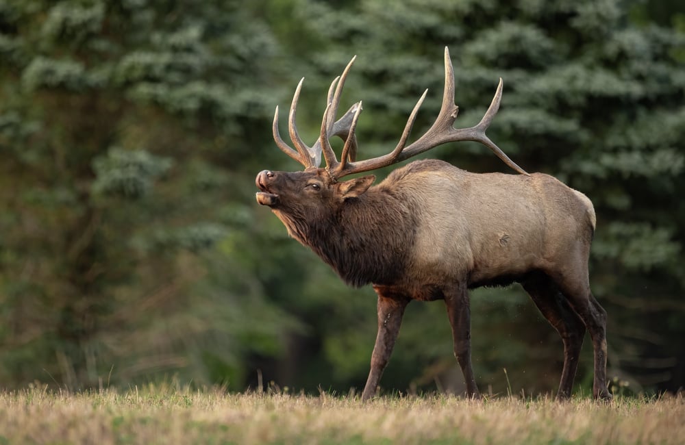
The elk (Cervus canadensis) is the second-largest cervid after the moose. Also called wapiti, these animals are occasionally confused with the moose, not only because they look similar, but because the moose is called “elk” in parts of Eurasia. The Roosevelt’s elk, Tule elk, Rocky Mountain elk, and the Manitoban elk are four of the six North American subspecies that still exist in the wild, while the Eastern and the Merriam’s Easter elk subspecies are extinct.1
Elk are related to an ancient breed of red deer that once lived in Asia. It is believed that they came to North America across the Bering Strait about 120,000 years ago, as did numerous other hoofstock species. Between 5,000 to 10,000 years ago, they advanced as far as Colorado, and there may have been as many as 10 million elk in North America when European settlement began.2
Elk are some of the most social of the cervids, with group sizes during the summer reaching up to 400 individuals. Adult males and females segregate themselves into different herds during most of the year, with female herds being larger and males creating small groups or being solitary.1,3 Younger bulls live in female herds or in groups of older, less aggressive bulls.
Male elk are called bulls and female elk are called cows. Male elk have antlers that are comprised of bone and grow about an inch a day. These can grow to over 20 inches in length. As they grow, a layer of velvet covers them, which is shed in the spring after the antlers are done developing. The North American and Siberian elk have the largest antlers, which can weigh up to 40lbs.1,2
Elk are browsers, feeding on grasses, sedges, and forbs in summer and woody growth during the winter months. They appear to be especially fond of dandelions, aster, hawkweed, violets, clover, and the occasional mushroom. Since elk are ruminants, they regurgitate their food and remasticate to aid in digestion.1,2
Shock and How it Arises
Shock is a critical condition that is caused by a sudden drop in blood flow throughout an animal’s body. It can be the result of a wide variety of conditions or circumstances, including extreme physical stress, trauma, disease, heatstroke, blood loss, allergic reactions or severe infection. When an animal is suffering from shock, its organs are not receiving an adequate amount of blood or oxygen. If untreated, this condition can lead to permanent organ damage or death.
Unfortunately, the processes surrounding capture and/or chemical immobilization can include extreme physical stress and/or trauma sufficient to induce shock in elk and other animals. The degree of risk is dependent upon factors such as sex, age, overall health, environmental factors, length of immobilization, the degree of stress involved in the capture/immobilization event itself, the specific chemical agents involved in immobilizing the animal and others. It should be noted that cervids are known to be susceptible to a variety of complications during chemical immobilization, including shock.
There are three main categories of shock:
Circulatory Shock. This occurs when there is a significant decrease in effective circulating blood volume. This category is further divided into the three subcategories of cardiogenic, hypovolemic and distributive shock. Cardiogenic shock occurs when the circulating volume of blood decreases despite normal or increased blood volume. Hypovolemic shock occurs when blood volume is decreased through hemorrhage, third space fluid distribution, or dehydration. Distributive shock occurs when an animal’s body is unable to maintain the vasoconstriction of blood vessels.5
The last two categories of shock are hypoxic shock and metabolic shock. Hypoxic shock results from impaired oxygen delivery to cells, while metabolic shock involves cells that have become unable to utilize oxygen for energy production.5,6For the purposes of this discussion, the types of shock being discussed are the subcategories of circulatory shock and hypoxic shock, which are the most likely to be brought on due to capture and/or chemical immobilization events.
The Mechanics of Shock in Elk
Even under well-controlled circumstances, chemical immobilization carries risks in elk. Almost all of the drugs that produce anesthesia compromise cardiovascular stability by producing dose-dependent impairment of cardiac function, vascular reactivity and autoregulatory responses.6 Hemoglobin is found within red blood cells and carries oxygen to tissues. In normal circumstances, the amount of oxygen delivered to cells is 2 to 4 times the amount required, which ensures an adequate supply.4 However, if tissues are not adequately perfused with blood, the oxygen fails to get to the cells, regardless of the oxygen content in the blood.5
Changes in the mean arterial pressure (MAP) will trigger changes in the heart rate.6,7 An increase in MAP also causes bradycardia and vasodilation, while a decrease produces tachycardia and vasoconstriction.5,6 While anesthesia-related depression of cardiac function and arterial vasodilation are adverse effects that are well-recognized as contributing to anesthetic risk, far less emphasis is usually placed on effects impacting venous physiology and venous return.4
Venous circulation represents approximately 70% of an animal’s total blood volume, and this is a chief contributor to stroke volume and cardiac output.5 Vasodilation is the primary cause of hypovolemia produced by anesthetic drugs. It is often associated with increased venous compliance, decreased venous return, and reduced response to vasoactive substances.6 Depending on things like patient status and monitoring, a state of relative hypovolemia can remain clinically undetected for long periods of time.5-7
Diagnosis and Treatment of Shock in Elk
The clinical signs of shock in elk can include any combination of the following symptoms:
- Unresponsiveness
- Hypothermia
- Tachycardia
- Bradycardia
- Tachypnea
- Bradypnea
- Marked hypotension
- Cyanosis
- Orthopnea
The treatment of shock in elk must focus primarily on increasing oxygen delivery to the tissues. This can be accomplished by providing supplemental oxygen, increasing effective circulating volume, increasing hemoglobin concentration and increasing cardiac output with stimulants.5,7 An intravenous catheter should be placed for vascular access if possible. If venous access cannot be established, an intraosseous catheter can be placed. Oxygen supplementation, when available, will also provide benefits to the elk suffering from shock. This can be accomplished via flow-by oxygen, mask, nasal cannulas or an oxygen cage.6
Fluid Therapy for Shock in Elk
Lactated Ringer’s solution, Normosol-R™, and Plasma-Lyte™ are the preferred choices for fluid therapy in treating shock in elk, as these have been shown to cause fewer complications as well as decreasing the risk of mortality5 as compared to other options. Hypertonic saline is also an accepted option for fluid therapy (as this increases vascular volume). Hypertonic saline increases plasma osmolarity, pulling water into the vascular space from the interstitial space, thereby expanding plasma volume. It should be noted that hypertonic saline does have unwanted side effects, such as a transient, dose-dependent increase in sodium and chloride.6,7
Blood products are an important adjunct for the treatment of shock in elk and other hoofstock. In normal patients, anemia can be well-tolerated with oxygen delivery being maintained. In patients with trauma and acute loss of blood volume however, the associated stressors can contribute to decreased oxygen delivery.7
1britannica.com.
2rmef.org.
3animaldiversity.org.
4todaysveterinarynurse.com.
5Noel-Morgan, J., Muir, W. (2018) Anesthesia-Associated Relative Hypovolemia: Mechanisms, Monitoring, and Treatment Considerations. Frontiers in Veterinary Science, Vol. 5 (53).
6Haller G, Laroche T, Clergue F. Morbidity in anaesthesia: today and tomorrow. Best Pract Res Clin Anaesthesiology (2011) 25(2):123–32.
7Steadman J, Catalani B, Sharp CR, Cooper L. Life-threatening perioperative anesthetic complications: major issues surrounding perioperative morbidity and mortality. Trauma Surg Acute Care Open (2017).


