One of the most dangerous conditions known to occur in wild and domestic animals is capture myopathy. This is described as muscle damage resulting from extreme exertion, struggle, or stress. It is also known as exertional myopathy, overstraining disease and exertional rhabdomyolysis.1 Capture myopathy most often occurs as a result of capture, transport or chemical immobilization, but it can also be the result of other natural causes of stress, such as in prey animals attempting to avoid or struggling with predator animals.2 Capture myopathy is of particular concern in cases when it is a cause of death in wild animals that are handled by humans.
Capture myopathy can occur in any animal under extreme physical stress, although it is thought that some species may be more predisposed to it than others due to their natural temperament and physiological characteristics.3 Capture myopathy has been most widely studied in ungulates and birds, although it is believed to potentially affect any captured wildlife species. It has also been observed in coyotes, badgers, primates, and many other species.2
Ungulates in North America that have been reported with this condition have included white-tailed deer, mountain goats, bighorn sheep, pronghorn antelope, bison, moose, and elk.2,6
Eld’s Deer: Background and Biology
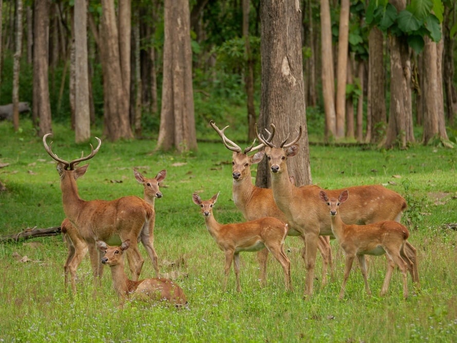
The Eld's deer (Rucervus eldii) is indigenous to Southeast Asia. It is a large deer that is considered very regal and graceful in appearance. These deer are similar in size to white-tailed deer, but differ in appearance. Their legs are long and thin, and they have slender bodies with a large head and ears. Their rough coats change color with the season. In summer, they are reddish-brown, and dark brown in winter. Stags often have darker coloring than hinds (females) and have a thick mane of long hair around the neck.4
Eld’s deer stags have large bow- or lyre-shaped antlers; these sweep back in a curve of about 40 inches in length, with one smaller tine growing toward the front of the head. Antlers are shed every year and reach their largest size during the breeding season.5 Male Eld’s deer grow to about 71 inches in length and weigh from 276 to 386 pounds. They are taller and larger than the hinds, which stand about 60 inches tall.
Rucervus eldii are primarily nocturnal deer. Throughout most of the year, stags tend to be loners, except in the spring when mating commences. Females are generally found alone or in pairs with their young. They remain in close association with their fawns and other female-fawn pairs. Larger groups are often formed when males join groups of females prior to the breeding season, and groups of up to 20 animals are common.4
In their native ranges, Eld’s deer inhabit suitable forest habitats, lowland valleys and plains, avoiding dense forests and coastal areas. This also includes monsoonal forests. Today, they occur in a number of protected areas throughout these areas and have been introduced to numerous countries as game animals, including the United States.4,5
Clinical Signs of Capture Myopathy in Eld’s Deer
Capture myopathy can occur naturally when a deer is attempting to avoid predation, but for the purposes of this discussion, capture myopathy will be the result of Eld’s deer being captured and/or immobilized, with or without chemical means involved. Deer are adapted to escape from predators, but they are not adapted to struggle for long periods of time in human-constructed restraints.3 When animals overexert themselves (e.g., struggling in a trap) to the extent that physiological imbalances develop and result in severe muscle damage, capture myopathy can result.2
Clinical signs of capture myopathy in Eld’s deer can vary depending on the cause of exertion.2 The method of capture and restraint is also a determinant in occurrences of capture myopathy. The available literature states that capture myopathy may result in sudden death, or that clinical signs may develop hours, days, or up to two months following capture.6 The clinical signs during early onset include elevated respiratory rate, heart rate, and body temperature.1,3 Body temperature increases during exertion, with higher temperatures being associated with death due to capture myopathy. The increase in body temperature can be above 42°C.4 Muscle spasms, stiffness and lameness are also clear signs of capture myopathy. Animals often become recumbent and may stumble. If dark red-colored urine is noted, this is an indication that the animal's muscles are breaking down and that its kidneys have been severely affected.2,6 Death of the animal usually follows. Upon necropsy, light-colored skeletal and cardiac muscle is indicative of capture myopathy being the cause of death.
Preventing Capture Myopathy in Eld’s Deer
Since there is no treatment for capture myopathy, prevention is the best method of avoiding this complication. Care should be taken in case of handling of animals that tend to be more susceptible to capture myopathy, which includes large hoofstock. An anesthetic protocol consisting of good anesthetic agents can aid significantly in preventing capture myopathy in deer. In the case of wild Eld’s deer, the remote delivery of anesthetic agents is considered a superior methodology to trapping prior to the anesthetic event.
The team in the field should be thoroughly aware of the risks of capture myopathy and make every effort to prevent its occurrence. Eld’s deer should only be captured when necessary, and the negative effects that capture may have on an animal's health should always be considered before beginning a capture or initiating an anesthetic event.7 Capture methods that minimize animal stress, struggling and handling time should be utilized.
Appropriate protocols for chemical immobilization may vary depending on the deer species, so research into the available recommendations for Eld’s deer may be helpful in identifying the ideal capture method. It has been reported that using a combination of Xylazine HCL and Ketamine HCL can decrease the chance of capture myopathy, but this is not a guarantee of avoiding capture myopathy in any deer.3
1Friend, M., Thomas, N. J. Field Manual of Wildlife Diseases. In: Field Manual of Wildlife Diseases, United States Geological Survey, 361-368.
2Williams, E. S., Thorne, E. T. 1996. Exertional Myopathy (Capture Myopathy). Noninfectious Diseases of Wildlife, Second Edition, 181-193 Iowa State University Press, Ames, Iowa, USA.
3Blumstein, D., et. al. The evolution of capture myopathy in hooved mammals: a model for human stress cardiomyopathy?Evolution, medicine, and public health vol. 2015,1 195-203. 21 Jul. 2015.
4nationalzoo.si.edu.
5animalia.bio.
6Kohn, Tertius. (2013). Capture myopathy mystery.
7Businga NK, Langenberg J, Carlson L. Successful treatment of capture myopathy in three wild greater sandhill cranes (Grus canadensis tabida). J Avian Med Surg. 2007 Dec;21(4):294-8. doi: 10.1647/2005-013R1.1. PMID: 18351009.


