The Eld’s deer (Rucervus eldii) is a tropical deer that is native to the lowland forests of an area known as the Indo-Burma Biodiversity Hotspot.1 This deer inhabits the forested areas, lowland valleys and plains of southeast Asia, avoiding dense forests and coastal areas. Today, Eld’s deer occur in several protected areas throughout their former range and have been introduced into numerous other countries as game animals.
Lt. Percy Eld discovered this species in the Manipur Valley of India in 1838, from which their name is derived. Due to rampant hunting and habitat conversion, populations of this deer have significantly declined in their native range and are now severely fragmented.
Three subspecies of Eld’s deer are currently recognized:
- The Sangai (Rucervus eldii eldii) of northeast India. This subspecies was once widespread in the Manipur Valley. It was once declared extinct, but was later rediscovered in Keibul Lamjao National Park.
- The Thamin (Rucervus eldii thamin) of Myanmar and western Thailand. The Thamin was once distributed widely in the central plains of Myanmar, but is now reduced to two major populations.
- The Siamese Eld’s Deer (Rucervus eldii siamensis) of Indochina and China’s Hainan Island, which once inhabited Thailand, Laos, Cambodia, Vietnam and Hainan. It was thought to be extinct in the wild across Indochina, but scattered populations were rediscovered in Laos and Cambodia in 1998. The Eld’s deer on China’s Hainan Island is the only island population of this species.2
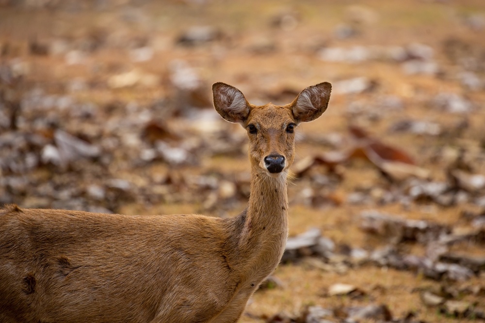
The Eld’s deer is a large deer that is considered regal and graceful in appearance. They are similar in size to white-tailed deer, but differ somewhat in body type. Their legs are long and thin, and they have slender bodies with a large head and ears. Their rough coats change color with the season. In summer, they are reddish-brown, and dark brown in winter. Stags often have darker coloring than hinds (females) and have a thick mane of long hair around the neck.1
Rucervus eldii are known to graze and browse on wild fruits and cultivated crops (such as rice, lentils, maize, peas and rape) from nearby fields. Rucervus eldii thamin has been reported to eat the fruits of various woody plant species. They also eat forbs and grasses in these areas.3 The Eld’s deer is closely associated with areas that are seasonally burned; they are often found eating new grasses as they emerge after a burn.1 Their feeding may vary seasonally with food availability and with reproductive considerations; during rut, males often experience a decline in body weight, probably due to a decrease in their food intake.
While Eld’s deer are usually solitary, they sometimes form large groups or herds as an anti-predator adaptation. These larger groups decrease the risk of predation, both by increasing the chance that a predator will target other animals rather than a lone individual, and by the increased vigilance for predators which can be provided by all members of the group.2The most common predators of Eld’s deer are tigers and leopards. Jackals and feral dogs occasionally hunt these deer, and poaching by humans is a serious problem to Eld's deer populations.1
Like most cervids, Eld’s deer mothers typically hide their fawns in underbrush immediately after birth. Females give birth during the cool-dry season when the flood waters have receded and vegetation has begun to grow, which provides the young with shelter and helps to conceal them.3 Fawns are weaned after 4 to 5 months, which allows them to have sufficient time to increase their mobility and ability to travel with the herd.1,2
Eld’s Deer, Chemical Immobilization and Aspiration Risks
Researchers and wildlife managers are often called upon to perform routine procedures on Eld’s deer, or to administer medical treatment. In most cases, this requires the use of sedative and/or anesthetic drugs. Vomiting is one of the more common post-sedation and post-anesthesia complications in both domestic and exotic animals. Vomiting that occurs during a procedure can pose grave risks due to anesthetic aspiration.
Aspiration is when a foreign substance enters the airway or lungs, which can apply to food, liquid, or other materials. Aspiration can give rise to serious health problems, such as aspiration pneumonia. Aspiration can occur when a human or animal has difficulty swallowing normally (which is referred to as dysphagia), but in some instances it can be brought on during or after anesthetic events, as indicated above.
Aspiration pneumonia is a condition that is characterized by inflammation and necrosis of lung tissue due to the inhalation of foreign material. The most common material aspirated in large animals is a large volume of liquid, due to weakness from a pathological condition, during oral administration of fluids or as a result of chemical immobilization. The severity of the inflammatory response will depend on the type and volume of material aspirated and the distribution of aspirated material in the lungs.
In cases in which large volumes of liquid have been aspirated, death can occur swiftly. Other cases will present later with clinical signs consistent with cranioventral bronchopneumonia.4 Cervids such as deer that are affected with chronic wasting disease may also develop aspiration pneumonia due to central nervous system dysfunction.3
Eld’s Deer and Chemical Immobilization
The field immobilization of wild animals with chemical agents is a method of rendering them tractable while using minimal restraint. Here, the research or wildlife management objectives are usually to measure or weigh the live deer, collection of blood or tissue for research or diagnostics, marking an individual or fitting a radio transmitter for studying migration patterns, range requirements and behavior patterns or the translocation of animals for a variety of reasons.4,5These requirements have resulted in the development of increasingly safer methods of chemical immobilization.
At the beginning of the last century, the primary method used for the capture of large wild animals such as deer was to chase them to the point of near-exhaustion, a time- and labor-intensive method that was impractical and fairly inhumane.4With the pioneering work on the chemical immobilization of wildlife that took place from the 1950s on, chemical immobilization techniques have improved greatly through the development of increasingly efficacious drugs and equipment.
Each species of deer has its own anesthesia recommendations with intra-species variations of dosages because of diverse individual responses to anesthetic agents.4,5 These variations are of course factors in the risk of vomiting and anesthetic aspiration in these species, and other factors (e.g., stress, venue, individual animal and field conditions) must also be taken into account.
Hyperthermia and capture myopathy are commonly-encountered problems with deer anesthesia. Intubation has been widely recommended for any anesthetized deer that needs to be transported or anesthetized for greater than one hour. Unfortunately, the literature strongly suggests that intubation can increase the risk of vomiting during an anesthetic event.4,5
Anesthetic Aspiration in Eld’s Deer
The utilization of basic veterinary knowledge can make a substantial contribution to animal safety during capture and chemical immobilization. Teams that are qualified to handle Eld’s deer should evidence the appropriate expertise in wildlife anesthesia and should include an attending veterinarian when appropriate. A successful chemical restraint exercise is not complete until the subject is fully recovered and (in the case of field operations) back in its environment. The application of appropriate pharmacological principles with an emphasis on drug reversibility will minimize the chances that the animal will be at a competitive disadvantage or inordinately disoriented following its release.4-6
Until formulated drugs (e.g., combinations of α2-agonists such as medetomidine, detomidine, xylazine and their reversal agents) came into use in recent years, opioids were the mainstay of large animal anesthesia in wildlife and captive care.5As with other mammals, problems encountered with certain opioids (such as etorphine or carfentanil, which have been widely used in wildlife chemical immobilization) in deer are known to include vomiting or passive regurgitation that can lead to aspiration pneumonia.
Periprocedural fasting (fasting prior to an anesthetic event) has historically been recommended by clinicians because of the suspected risk of aspiration. Unfortunately, this is very often impossible under field conditions. Additionally, much of the data on anesthetic aspiration relates to humans receiving general anesthesia, however, Eld’s deer and other mammals have been known to aspirate during procedures while under sedation and where no intubation or general anesthesia were employed.
For the prevention of anesthetic aspiration, the literature recommends histamine (H2) antagonists such as cimetidine, famotidine, nizatidine, and ranitidine and proton pump inhibitors (PPIs) such as dexlansoprazole, esomeprazole, lansoprazole, omeprazole, pantoprazole, and rabeprazole, which have been shown to be effective in increasing the pH and reduce the volume of gastric contents.5 Prokinetics (e.g., domperidone, metoclopramide, erythromycin and renzapride) promote gastric emptying and are also believed to reduce the risk of aspiration.6
In the event that aspiration occurs during a procedure in an Eld’s deer, the first step in managing the situation is the recognition of gastric content in the oropharynx or the airways.5-8 The deer should be immediately positioned with the head down and rotated laterally if possible. Orotracheal and endotracheal suctioning should be performed, either before or after orotracheal intubation, depending on whether regurgitation continues and if the airway is visible.
The airway should be secured as rapidly as possible to prevent further contamination and to facilitate airway clearance.5Flexible bronchoscopy may be used, as this is an important adjunct to orotracheal and endotracheal suctioning. Rigid bronchoscopy may be required if particulate matter is present in the deer’s airway.4,7
1animaldiversity.org.
2nationalzoo.si.edu.
3animalia.bio.
4Merck Veterinary Manual.
5Shaikh, Safiya Imtiaz et al. Postoperative nausea and vomiting: A simple yet complex problem. Anesthesia, essays and researches vol. 10, 3 (2016).
6Nason, K. Acute Intraoperative Pulmonary Aspiration. Thoracic surgery clinics vol. 25,3 (2015): 301-7.
7Lance, W. Exotic Hoof Stock Anesthesia and Analgesia: Best Practices. In: Proceedings, NAVC Conference 2008, pp. 1914-15.
8Kluger M.T., et. al. Crisis management during anaesthesia: regurgitation, vomiting, and aspiration. Quality & safety in health care. 2005;14(3): e4.


